
Fig. 2
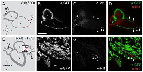
Isl1 expression in the embryonic and adult zebrafish heart. Single confocal scans of a fluorescent antibody labeling of Isl1 and eGFP in embryonic (2 dpf) (A?D) and adult (E?H) zebrafish expressing Tg(myl7:eGFP) in all cardiomyocytes. GFP+ cardiomyocytes are displayed in grey (B, F) and in green in (D, H). Isl1 is shown in grey (C, G) and in red (D, H). Arrowheads indicate Isl+/GFP+ cells. Illustrations of a lateral view of a 2 dpf (A) and adult (E) zebrafish heart indicate the location of Isl1+ cells (red). The box in panel E represents the area shown in (F?H). (B?D) Fluorescent immunolabeling of Isl1 and eGFP in a 2 dpf embryo (sagittal section 100 μm). At this time point Isl1+/GFP+ cells were only found in the IFT of the heart. (F?H) Fluorescent immunolabeling of Isl1 and eGFP in an adult zebrafish heart (sagittal section 100 μm). Isl1+/GFP+ cells are located at the junction of the sinus venosus and atrium in the inflow region of the heart (arrowheads). Isl1+ cells showed low expression of myl7. v, ventricle; a, atrium; oft, outflow tract; ift, inflow tract; ba, bulbus arteriosus; sv, sinus venosus; a, anterior; p, posterior; d, dorsal; v, ventral. Scale bars represent 50 μm. |
| Gene: | |
|---|---|
| Antibody: | |
| Fish: | |
| Anatomical Terms: | |
| Stage Range: | Long-pec to Adult |