
Fig. 2
- ID
- ZDB-FIG-121116-29
- Publication
- Robu et al., 2012 - Rereplication in emi1-Deficient Zebrafish Embryos Occurs through a Cdh1-Mediated Pathway
- Other Figures
- All Figure Page
- Back to All Figure Page
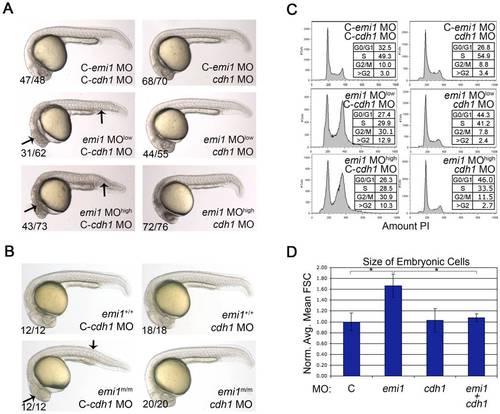
Cellular and developmental defects caused by emi1 knockdown are due to cdh1 activity. (A and B) Phase-contrast analysis of embryos at 24 hpf. (A) Morpholino injected embryos, where C- indicates the mismatch control morpholino for the indicated gene. We injected 1 ng of control and cdh1 morpholinos per embryo. The emi1 morpholino was injected at 1 ng (low) or 2.7 ng (high) per embryo. (B) Genotyped wild-type siblings (+/+) and homozygous emi1 mutant embryos (m/m) injected with 2 ng control or cdh1 morpholinos as indicated. Note the wild-type appearance of homozygous mutants and emi1 morphants that were injected with cdh1 morpholino. (C) Cell cycle analysis by FACS scanning of propidium iodide (PI)-stained cells. Total cells were analyzed from single cell suspensions of pools of the indicated morpholino-injected embryos at 4–5 somites. A representative experiment is shown. (D) The average cell size from FACS analysis of cells from morpholino-injected disaggregated embryos at 4–5 somites, normalized to the average control cell size (C, arbitrarily set at 1.00). Error bars indicate standard deviation (SD). Cell size data and SD were obtained from 3 independent experiments. |
| Fish: | |
|---|---|
| Knockdown Reagents: | |
| Observed In: | |
| Stage Range: | 1-4 somites to Prim-5 |