
Fig. S4
- ID
- ZDB-FIG-120907-32
- Publication
- Salbreux et al., 2012 - Coupling mechanical deformations and planar cell polarity to create regular patterns in the zebrafish retina
- Other Figures
- All Figure Page
- Back to All Figure Page
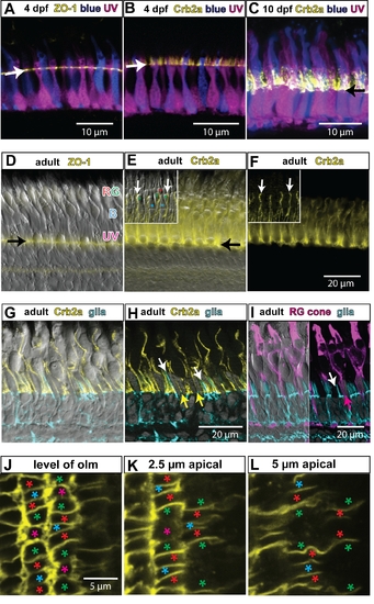
Differentiation of cone photoreceptors and elaboration of the apical process. A) Retinal cryosection from larval transgenic (mi2009) fish at 4 days post-fertilization (dpf), in which blue and UV cones express the reporters mCherry and EGFP (pseudocolored blue and magenta, respectively), which fill the cytoplasm of the cells. The OLM (arrow) is labeled by ZO-1 immunostaining (yellow). The developing inner and outer segments of the cones project apically beyond the OLM. B) Larval mi2009 fish at 4 dpf immunolabeled for Crb2a (yellow). Note that the Crb2a protein extends beyond the OLM (arrow) on the plasma membrane of the inner segments. C) By 10 dpf, the cone inner and outer segments have elongated further and the Crb2a protein also extends further apically on the inner segments. The arrow indicates the level of the OLM. D) Differential interference contrast (DIC) image of a retinal cryosection from an adult zebrafish, immunolabeled with ZO-1 (yellow) to label the OLM (arrow). The inner and outer segments of cones extend apically; the UV cones are the shortest, the blue (B) cones are longer, and the red and green (RG) double cones are the longest. E) A DIC image with immunolocalization of Crb2a protein on the inner segments of the cones. The black arrow indicates the OLM. The inset shows the Crb2a protein at the interface between red and green double cones (white arrows). F) Same image as panel E without the DIC channel. G) Differential interference contrast (DIC) image of a retinal cryosection from an adult transgenic zebrafish (mi2002), expressing a fluorescent reporter in Müller glia (cyan), and immunolabeled with Crb2a (yellow). H) Same image as panel E without the DIC channel. The processes of Müller glia (white arrows) extend apically beyond the OLM, but not as far as the Crb2a (yellow). Müller processes do not separate the interface between the inner segments of red and green double cone pairs, which have strong staining for Crb2a (yellow arrows). I) Left half: DIC image of a retinal cryosection from an adult transgenic zebrafish (mi2002), expressing a fluorescent reporter in Müller glia (cyan), and immunostained with zpr1, which labels red and green cones (magenta). Right half: same image without the DIC channel. The processes of the Müller glia (white arrow) are not interposed between the inner segments of red and green double cone pairs (magenta arrow). J–L) Individual optical sections from a z-stack confocal image of Crb2a immunolabeling near the retinal margin in a retinal flat-mount (cropped version from the same image series shown in Figure 6A). Panel J is at the level of the OLM, panel K is the subapical region (SAR) at 2.5 μm from the OLM, and panel L is the SAR at 5 µm from the OLM. Identity of cone photoreceptor subtypes is indicated by asterisks: red, green, blue, and UV (magenta), respectively. In the SAR, Crb2a has a planar polarized distribution – it is expressed at higher levels at the interfaces of red, green, and blue cones within a column. The retinal surface is curved, so the left and right sides of each panel are more apical than the center (and thus show a somewhat more polarized Crb2a distribution in J and K). |