
Fig. 6
- ID
- ZDB-FIG-120720-16
- Publication
- Harden et al., 2012 - Close association of olfactory placode precursors and cranial neural crest cells does not predestine cell mixing
- Other Figures
- All Figure Page
- Back to All Figure Page
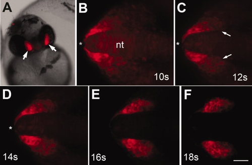
Six4b:mCherry expression starts during somitogenesis. A: Expression in olfactory placodes (OPs) in founder at 48 hours postfertilization (hpf). B–F: Stills from 6-hr time lapse generated starting at 10s (see Supp. Movie S1). B–F: The OP precursors are concentrated in the most rostral region of the head (asterisk). C–F: The mCherry-expressing cells located at the posterior edge of the OP field (C, arrows) coalesce, forming the posterior border (D–F). B–D: During this time, the anterior mCherry-expressing cells move caudally away from the tip of the neural tube (asterisks) aggregating to form the olfactory placodes (E,F). All images are dorsal views with rostral to the left. nt, neural tube. Scale bar = 30. (See Supp. Movie S2, S3.) |