FIGURE

Fig. S4

ID
ZDB-FIG-120612-21
Publication
Lindeman et al., 2012 - Localized Products of futile cycle/ lrmp Promote Centrosome-Nucleus Attachment in the Zebrafish Zygote
Other Figures
All Figure Page
Back to All Figure Page
Fig. S4

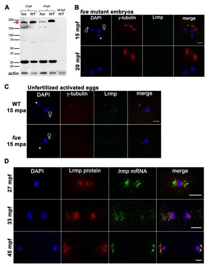
Western Blotting and Additional Protein/RNA Localization Analysis, Related to Figure 4

(A) Embryo lysates from wild-type and two fue mutant clutches at cleavage stages and 4 hpf were probed with anti-LrmpMD antibody. A band at 200 kDa (red arrow) likely corresponds to Lrmp and appeared reduced in mutant compared to wild-type, while a shorter 160 kDa band was present in mutant lysates. The observed 200 kDa molecular weight is greater than predicted for the zebrafish Lrmp isoforms (approximately 159 kDa - 162 kDa), however mouse and human Lrmp proteins also exhibit higher-than-expected molecular weights on denaturing gels (predicted at 62 kDa and 59 kDa, observed at 75 kDa and 69 kDa, respectively) [11,44]. The identity of the 25 kDa band seen in both mutant and wild-type is not known and unlike the 200 kDa species, was not recognized by the more C-terminal Lrmp antibody (data not shown). Wild-type 24 hpf lysates did not show any prominent signal. Anti-actin antibody was used as a loading control.
(B) fue embryos were labeled with DAPI (blue), γ-tubulin antibody (red) and anti-LrmpMD antibody (green). Localized Lrmp protein was undetectable in the majority of imaged fue embryos (16 out of 20), and only very faint nuclear envelope labeling could be seen in the remaining mutants (4 of 20).
(C) In unfertilized activated wild-type eggs at 15 minutes post-activation (mpa), Lrmp protein is localized at the female pronuclear membrane at levels comparable to wild-type zygotes (top row; compare to Figure 4, top three rows). Similar to fertilized fue zygotes, localized Lrmp protein is not readily detectable in unfertilized fue eggs (bottom row).
(D) Fluorescent in situ labeling against lrmp mRNA (green) in combination with antibody labeling for Lrmp protein (red) in wild-type embryos. In early mitosis, Lrmp protein and lrmp mRNA are offset with Lrmp protein closer to the condensing DNA (top row). During chromosome segregation at anaphase, lrmp mRNA and Lrmp protein colocalize in spindle regions but only Lrmp protein appears at the reforming nuclear membranes (second row). Lrmp protein alone continues to localize at nuclear membranes in late mitosis, when Lrmp protein labeling at centrosomes again becomes pronounced, co-localizing with lrmp mRNA in this structure (bottom row in (D)). In (B,C), male and female pronuclei are indicated with symbols while polar bodies are denoted by white asterisks. Scale bars represent 20 μm.

Expression Data

Expression Detail
Antibody Labeling
Phenotype Data

Phenotype Detail
Acknowledgments
This image is the copyrighted work of the attributed author or publisher, and ZFIN has permission only to display this image to its users. Additional permissions should be obtained from the applicable author or publisher of the image. Full text @ Curr. Biol.