
Fig. 2
- ID
- ZDB-FIG-120522-34
- Publication
- Fleming et al., 2004 - A central role for the notochord in vertebral patterning
- Other Figures
- All Figure Page
- Back to All Figure Page
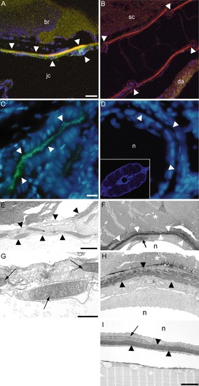
Ossification in the zebrafish vertebral column. (A,B) Simultaneous labelling at 20 dpf with alizarin red (red) for bone, zns5 (green) for osteoblasts, and DAPI (blue) for nuclei. (A) Confocal microscopic image of a sagittal section through the intramembranous parasphenoid bone (arrowhead) separating the jaw cavity (jc) from the brain (br). Osteoblasts in bone matrix are indicated by co-localization of alizarin red and zns5 (yellow). (B) Representative confocal microscopic image of a sagittal section through a thoracic centrum showing bone (red) but not zns5 labelling, indicating the absence of osteoblasts (all centra were examined in 10 serially sectioned embryos). Arrowheads indicate centrum boundaries. da, dorsal aorta; sc, spinal cord. (C,D) Osteoblasts assessed by alkaline phosphatase activity (green) using wide-field fluorescent microscopy. DAPI (blue) labels cell nuclei. (C) Representative transverse section through the epiphyseal bar of the skull (arrowheads) at 20 dpf showing large numbers of osteoblasts. (D) Representative transverse section (20 µm) through a mid-trunk centrum at 20 dpf, with no osteoblast labelling in the notochord (n) and surrounding centrum (arrowheads); inset shows a view at ~0.1× (all centra were examined in 10 serially sectioned embryos). (E-H) Analysis of bone by transmission electron microscopy (20 dpf). (E) Intramembranous ossification in the parasphenoid bone; osteoblast nuclei are clearly seen within bone matrix (arrowheads). (F) A centrum in transverse section showing acellular ossification. Bone matrix (arrowheads) lies adjacent to muscle (asterisk), encircling the notochord (n). In all samples (15 sections, 5 embryos) no osteoblasts were found. A notochord cell nucleus (arrow) lies adjacent to the matrix on the inner surface of the centrum. (G,H) High magnification images of E and F, respectively. Cell nuclei (arrows) are conspicuous within the intramembranous bone (G) but not centrum matrix (H). The latter is electron dense and lamellated (arrowheads). (I) Centrum in sagittal section showing bone matrix (arrowheads) lacking osteoblasts; the gap between bone and muscle (asterisk) is due to shearing during processing; arrow marks a nucleus from the notochord (n). Scale bars: A,B, ~10 µm; C,D, ~5 µm; E,F,~ 2 µm; G,H, ~1 µm; I, ~4 µm. |
| Antibody: | |
|---|---|
| Fish: | |
| Anatomical Term: | |
| Stage: | Days 14-20 |