
Fig. 5
- ID
- ZDB-FIG-120508-69
- Publication
- Zhang et al., 2012 - Laser ablation of the sonic hedgehog-a-expressing cells during fin regeneration affects ray branching morphogenesis
- Other Figures
- All Figure Page
- Back to All Figure Page
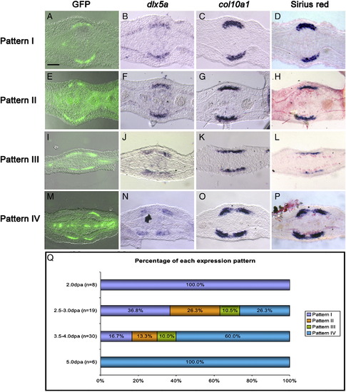
Separation of GFP domain of expression into two precedes separation of the domains of expression of bone markers and bone matrix. Representative examples of the four patterns of expression of GFP (A, E, I and M), dlx5a (B, F, J and N), col10a1 (C, G, K and O) observed in consecutive transverse sections of fin rays collected between 2 and 5 dpa. (D, H, L and P) Picrosirius red staining of the sections shown in (C, G, K and O), respectively. (Q) Percentages of fin rays showing pattern I (A–D), pattern II (E–H), pattern III (I–L), and pattern IV (M–P) at 2.0 dpa, 2.5–3.0 dpa, 3.5–4.0 dpa and 5 pda. Scale bar (all panels): 20 μm. |
Reprinted from Developmental Biology, 365(2), Zhang, J., Jeradi, S., Strähle, U., and Akimenko, M.A., Laser ablation of the sonic hedgehog-a-expressing cells during fin regeneration affects ray branching morphogenesis, 424-433, Copyright (2012) with permission from Elsevier. Full text @ Dev. Biol.