
Fig. S3
- ID
- ZDB-FIG-120423-48
- Publication
- Boggetti et al., 2012 - NBP, a zebrafish homolog of human Kank3, is a novel Numb interactor essential for epidermal integrity and neurulation
- Other Figures
- All Figure Page
- Back to All Figure Page
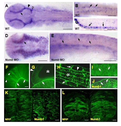
Distribution of NBP transcripts revealed by whole-mount in situ hybridization in wild-type (A-C) and Numb MO (D,E) embryos (all embryos are at 20-somite stage except for the 24 hpf embryo in C). (A,B,D) Flat preparations (anterior to the left), (C) section of the trunk region, (E) lateral view (anterior to the left). (A) NBP transcripts were ubiquitously detected and more pronounced in cells surrounding dorsally the eyes (arrow), at the ventral midbrain (filled arrowhead) and at the midbrain hindbrain boundary (open arrowhead). (B) Detail of the trunk region where NBP was also visible in few cells in each somite (arrows). (C) At 24 hpf, NBP was clearly concentrated at the epidermis (arrows point to the positive basal layer of keratinocytes). (D,E) NBP expression in Numb morphants presented a more disperse and expanded pattern in the head (D, compare with A) and in the trunk (E, compare with Fig. 2F) than in wild-type. NBP-GFP fusion protein localization (F-I). In 24 hpf embryos, NBP is cytoplasmic in neural tube cells (star in H), retinal cells (arrowheads in F) and otic epithelial cells (asterisk in G shows the otic vesicle). Over-accumulation of the fusion protein was observed at the basal pole of the cells at their attachments with the basement membrane (arrowhead in H points to the basement membrane surrounding the neural tube, arrows in H point to somitic boundaries to which muscle fibers anchor, arrows in F point to the inner basement membrane to which the retinal cells are attached, arrow in G points to the basement membrane between the otic vesicle and the hindbrain, arrowhead in G points to the basement membrane of the otic vesicle). In epidermal cells, NBP-GFP fusion is accumulated basolaterally (I shows tangential section, arrows point to the lateral membrane) similarly to Numb1 (arrows in J; this sample was fixated, embedded and probed with anti-HA antibody as in Supplemental Fig. S3K and Fig.2N). Immunodetection of NBP and Numbs (K,L; proteins were localized separately). The antibodies against the HA epitope (for Numbs) and c-myc epitope (for NBP) were growing in mouse and secondary antibodies were conjugated with Alexa 488. NBP and Numb1 when co-expressed were localized exclusively at the cell periphery (K). When NBP was co-expressed with Numb3, both proteins were distributed through the whole cells (L). Scale bar: A,D=100 μm, B,C,E=50 μF-L=10 μm. |
Reprinted from Developmental Biology, 365(1), Boggetti, B., Jasik, J., Takamiya, M., Strähle, U., Reugels, A.M., and Campos-Ortega, J.A., NBP, a zebrafish homolog of human Kank3, is a novel Numb interactor essential for epidermal integrity and neurulation, 164-174, Copyright (2012) with permission from Elsevier. Full text @ Dev. Biol.