|
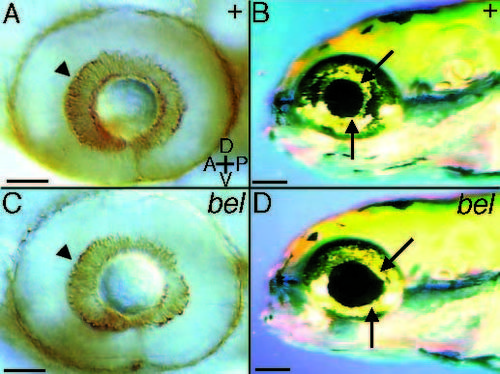
belladonna mutant eye phenotype. Wild-type (A) and belladonna (C) eyes at 48 hours labeled with the ZN-5 antibody to visualize retinal ganglion cell bodies. The retinal ganglion cells (arrowheads) and the entire eye appear normal at this age, thus the mutation does not appear to affect RGC differentiation. At 5 days iridophores and the pigmented epithelium are adjacent to the lens in the wild type (B). In belladonna mutants (D), gaps appear between the lens and some parts of the pigmented epithelium (arrow). The lens appears normal however. A, anterior; P, posterior; D, dorsal; V, ventral. Scale bars, 50 µm (A,C), 100 µm (B,D).
|