
FIGURE
Fig. 6
Fig. 6
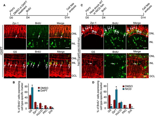
Notch Signaling Regulates the Differentiation of MG-Derived Progenitors (A and C) Diagram of the experimental protocol and representative photomicrographs showing BrdU and retinal cell type-specific immunofluorescence. Arrows identify BrdU and retinal cell type double-labeled cells. Scale bars, 50 μm. ONL, outer nuclear layer; INL, inner nuclear layer; GCL, ganglion cell layer. (B and D) Quantification of double-labeled cell types. *p < 0.01. Error bars represent SD. |
Expression Data
Expression Detail
Antibody Labeling
Phenotype Data
Phenotype Detail
Acknowledgments
This image is the copyrighted work of the attributed author or publisher, and
ZFIN has permission only to display this image to its users.
Additional permissions should be obtained from the applicable author or publisher of the image.
Reprinted from Developmental Cell, 22(2), Wan, J., Ramachandran, R., and Goldman, D., HB-EGF Is Necessary and Sufficient for Müller Glia Dedifferentiation and Retina Regeneration, 334-347, Copyright (2012) with permission from Elsevier. Full text @ Dev. Cell