
Fig. S11
- ID
- ZDB-FIG-120202-61
- Publication
- Dohn et al., 2012 - Distinct phases of Wnt/β-catenin signaling direct cardiomyocyte formation in zebrafish
- Other Figures
- All Figure Page
- Back to All Figure Page
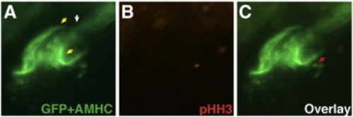
pHH3 marked proliferating cell in a zebrafish heart. Representative heart from Tg(hsp70l:wnt8a-GFP)w34;Tg(kdrl:nlsEGFP)zf109bv embryo stained for pHH3 (red) and S46 (AMHC; green) at 26 hpf after heat-shock at 16s. (A) Nuclear GFP marking the endothelial cells (white arrow) is internal to the AMHC (yellow arrows) expressing atrial myocardial cells. (B) A single cell is stained for pHH3. (C) Overlay indicating that the single cell positive for pHH3 (red arrow) is an endothelial cell. There was not a difference in the number of embryos with pHH3 positive myocardial cells or pHH3 positive cells per embryo (see Supplemental Tables 5 and 6). pHH3 marked proliferating cell in a zebrafish heart. Representative heart from Tg(hsp70l:wnt8a-GFP)w34;Tg(kdrl:nlsEGFP)zf109bv embryo stained for pHH3 (red) and S46 (AMHC; green) at 26 hpf after heat-shock at 16s. (A) Nuclear GFP marking the endothelial cells (white arrow) is internal to the AMHC (yellow arrows) expressing atrial myocardial cells. (B) A single cell is stained for pHH3. (C) Overlay indicating that the single cell positive for pHH3 (red arrow) is an endothelial cell. There was not a difference in the number of embryos with pHH3 positive myocardial cells or pHH3 positive cells per embryo (see Supplemental Tables 5 and 6). |
Reprinted from Developmental Biology, 361(2), Dohn, T.E., and Waxman, J.S., Distinct phases of Wnt/β-catenin signaling direct cardiomyocyte formation in zebrafish, 364-76, Copyright (2012) with permission from Elsevier. Full text @ Dev. Biol.