
Fig. 1
- ID
- ZDB-FIG-111202-12
- Publication
- Kizil et al., 2011 - Cerebroventricular Microinjection (CVMI) into Adult Zebrafish Brain Is an Efficient Misexpression Method for Forebrain Ventricular Cells
- Other Figures
- All Figure Page
- Back to All Figure Page
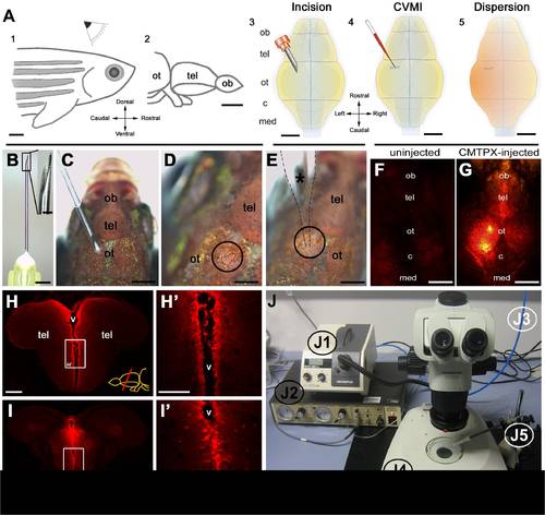
Overview of cerebroventricular microinjection (CVMI) paradigm and its target regions. (A) CVMI is performed at the dorsal surface of the head (1) and it targets, in this example, the forebrain that is rostral to the optic tectum (2). For injection, an incision is made into the skull over the optic tectum using a barbed-end canula (3). Through this slit, liquid is injected using a glass capillary (4). Injected liquid disperses rostrally (5). (B) The canula used for incision. (C) The incision on an adult fish. Dorsal view. (D) The incision site marked by dashed lines. (E) Injection with the glass capillary (*) (dotted lines mark the outline). (F) Dorsal view of an uninjected adult zebrafish head in red fluorescence channel. (G) Dorsal view of a CMTPX-injected adult zebrafish head in red fluorescence channel. (H) Cross section through the telencephalon. Ventricular cells are labelled with CMTPX. (H′) Higher magnification of the box in H. (I) Cross section through the midbrain. Ventricular cells are labelled with CMTPX. (I′) Higher magnification of the box in I. (J) Injection apparatus. J1: halogen light source with ring illuminator. J2: vacuum pump for microinjection. J3: pressurized air source. J4: dissecting microscope. J5: injection holder, needle and tubing. Scale bars: 500 μm A–G, 100 μm H-I′. ot: optic tectum, tel: telencephalon, ob: olfactory bulb, c: cerebellum, med: medulla, v: ventricle. |