
Fig. 1
- ID
- ZDB-FIG-111026-1
- Publication
- Smith et al., 2011 - Bmp and Nodal Independently Regulate lefty1 Expression to Maintain Unilateral Nodal Activity during Left-Right Axis Specification in Zebrafish
- Other Figures
- All Figure Page
- Back to All Figure Page
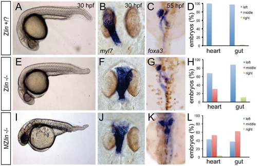
Dorsoventral and laterality defects in zygotic and maternal zygotic lin mutant embryos. (A–D) Wild-type zygotic lin siblings with normal ventral tail fin (A), left-positioned heart tube (B) and normal organ situs with liver on the left, pancreas on the right and left looped gut tube (C). (D) Quantification of heart position (n = 14) and direction of gut looping (n = 40). (E–H) Zygotic lin (Zlin) mutant embryos displayed a mild reduction of the ventral tail fin (n = 100/108) (E). In addition, in almost 30% of Zlin mutant embryos, the heart tube was positioned at the midline (F). Gut laterality was unaffected in Zlin mutant embryos (G). Quantification of heart position (n = 108) and direction of gut looping (n = 100). (I–L) Maternal zygotic lin (MZlin) mutant embryos derived from a cross of a homozygous lin mutant female and male showing the more severe posterior truncation (I) compared to a Zlin mutant embryo (E). In addition, most MZlin mutant embryos displayed a laterality defect in the heart (J), liver (bilateral, K) and in looping of the gut (K). (L) Quantification of heart positioning (n = 151) and direction of gut looping (n = 16). |
| Fish: | |
|---|---|
| Observed In: | |
| Stage Range: | Prim-15 to Day 5 |