
Fig. 4
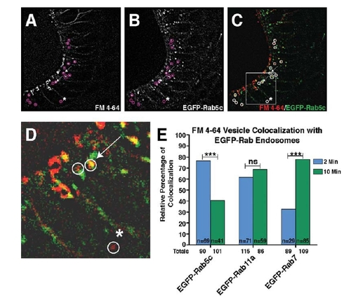
FM 4-64 injections into the developing otic vesicle indicate progression of endosome sub-types. A–C: 50-µm images thresholded for high fluorescence providing an example of FM 4-64 dye injection into the otic vesicle of a 28hpf EGFP-Rab5c embryo, 2 min post-injection. Single-channel images show intracellular puncta positive for the FM 4-64 dye (A), EGFP-Rab5c (B), and merged in C. D: Boxed image in C is magnified. Circles in A–D indicate non-membrane-associated, intracellular FM 4-64-positive vesicles. Arrows indicate examples of vesicles positive for both EGFP-Rab5c and the FM 4-64 dye, while the asterisk (*) indicates an FM 4-64-positive vesicle that is EGFP-Rab5c negative. E: Quantification of the percentage of FM 4-64-positive vesicles that co-localize with EGFP-Rab-positive vesicles at 2 and 10 min post-FM 4-64 dye injection. Numerical values below the x-axis indicate total number of FM 4-64 puncta analyzed from a minimum of 10 embryos. n-values indicate total number of FM 4-64 vesicles positive for EGFP-Rab at each time-point. Statistical Analysis: Fisher′s Exact Test. ***P < 0.001; ns, not significant. |