
FIGURE
Fig. 2
- ID
- ZDB-FIG-111011-5
- Publication
- Stevens et al., 2011 - Plasticity of photoreceptor-generating retinal progenitors revealed by prolonged retinoic acid exposure
- Other Figures
- All Figure Page
- Back to All Figure Page
Fig. 2
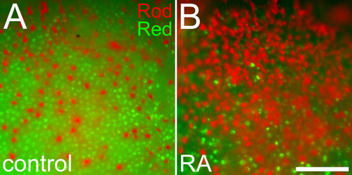
Altered positioning of rods in relation to red cones in response to retinoic acid treatment. Embryos treated with DMSO (A) or 0.3 μM RA (B) at 36 hpf and fixed at 60 hpf were doubly hybridized as whole mounts with probes corresponding to rod opsin (red color) and red cone opsin (green color). Following RA treatment, rods are more abundant, cones are more sparsely distributed, and rods display spacing characteristics more typical of cones. Bar = 50 μm. |
Expression Data
Expression Detail
Antibody Labeling
Phenotype Data
Phenotype Detail
Acknowledgments
This image is the copyrighted work of the attributed author or publisher, and
ZFIN has permission only to display this image to its users.
Additional permissions should be obtained from the applicable author or publisher of the image.
Full text @ BMC Dev. Biol.