
Fig. 1
- ID
- ZDB-FIG-110914-17
- Publication
- Ding et al., 2011 - Zebrafish as a potential model organism for drug test against hepatitis C virus
- Other Figures
- All Figure Page
- Back to All Figure Page
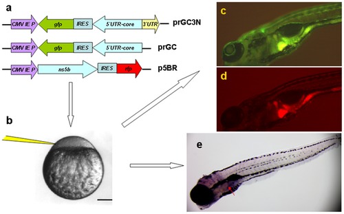
Zebrafish as a model organism for HCV sub-replicon amplification. a. The HCV sub-replicon was created with prGC3N and p5BR vectors. Cartoons are for vector prGC3N, prGC and p5BR. CMV promoter was for transcription. prGC3N has a gfp-IRES(EMCV)-core-52UTR sequence that was reversely inserted at the downstream of CMV promoter and followed by a HCV 32UTR sequence in a forward direction; prGC has no HCV 32UTR; the p5BR is a functional vector carrying HCV RNA polymerase (NS5B) and RFP. b. Co-injection of the sub-replicon into zebrafish zygote blastomere. c. Fluorescent microscopy examination for HCV core protein amplification in zebrafish liver using a GFP filter (480 nm excitation, 505 nm emission; image, 100X). d. Fluorescent RFP filter (556 nm excitation, 586 nm emission) was used to detect liver HCV NS5B protein signal in red. e. The whole mount in situ nucleic acid hybridization was used to detect the positive strand of HCV core RNA, in order to confirm the green fluorescent signal for the replication of the HCV sub-replicon. |