
Fig. 11
- ID
- ZDB-FIG-110706-23
- Publication
- Budi et al., 2011 - Post-embryonic nerve-associated precursors to adult pigment cells: genetic requirements and dynamics of morphogenesis and differentiation
- Other Figures
- All Figure Page
- Back to All Figure Page
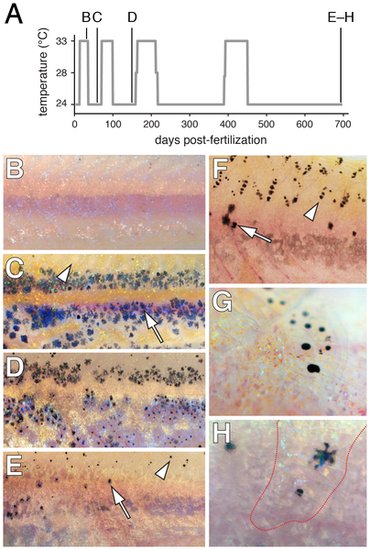
Limited regeneration of adult hypodermal melanophores following genetic ablation. (A) Time-course of temperature shifts, with letters corresponding to images in B–H. Final sample size at 698 days post-fertilization: n = 5. (B) A young adult kita; csf1rTS mutant at 33°C lacked melanophores as in kita; csf1rj4e1 mutants [38] (some melanized cellular debris resulting from melanophore death is evident dorsally). (C) Temperature downshift to 24°C allowed recovery of a hypodermal melanophore (e.g., arrow) complement initially indistinguishable from kita single mutants [59]. Arrowhead, the dorsal flank is initially devoid of scale-associated melanophores, as is typical of kita mutants. (D,E) Additional rounds of ablation and recovery yield progressively fewer hypodermal melanophores (arrow), though some melanophores develop on the dorsal scales (arrowhead). Hypodermal xanthophores and iridophores were depleted as well (data not shown). (F) Dorsal flank of another individual showing hypodermal melanophores (arrow) and scale melanophores (arrowhead). (G) Detail of scale-associated melanophores. Iridescent iridophores and yellow-orange xanthophores are apparent as well. (H) Detail of hypodermal melanophores viewed through an overlying scale containing a concentration of xanthophores and iridophores (outlined in red). |