
Fig. 3
- ID
- ZDB-FIG-110624-33
- Publication
- Rothenaigner et al., 2011 - Clonal analysis by distinct viral vectors identifies bona fide neural stem cells in the adult zebrafish telencephalon and characterizes their division properties and fate
- Other Figures
- All Figure Page
- Back to All Figure Page
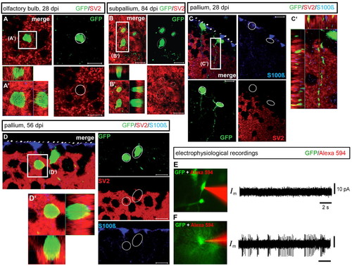
Structural and functional integration of adult-born neuron into existing forebrain neuronal networks. (A-D2) Immunohistochemical detection of GFP (green), S100β (blue) and the synaptic marker SV2 (red) on cross-sections of the OB (A,A2), subpallium (B,B2) or pallium (C-D2) at 28, 56 or 84 dpi (confocal z-section images and orthogonal projections). Scale bars: 10 μm. (E,F) Cell-attached electrophysiological recordings of GFP-positive cells. (E) Example of a GFP-positive cell 2 weeks post-infection that did not exhibit spontaneous action potentials. (F) Example of a GFP-positive cell 7 weeks post-infection with spontaneous action potentials (green, GFP-positive cells; red, contents of the patch pipette). Both right-panel horizontal and vertical scale bars are the same in E and F. |