FIGURE
Fig. 1
- ID
- ZDB-FIG-110608-20
- Publication
- Tu et al., 2011 - Fate restriction in the growing and regenerating zebrafish fin
- Other Figures
- All Figure Page
- Back to All Figure Page
Fig. 1
|
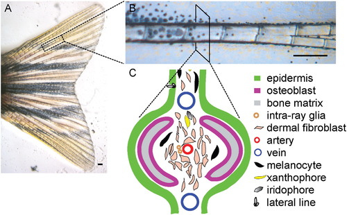
The Anatomy and Different Cell Types of the Zebrafish Caudal Fin (A–C) The zebrafish adult caudal fin is almost transparent, except that it has some pigmented cells: the melanocytes (black cells), xanthophores (yellow cells), and iridophores (shiny cells) (A). The caudal fin is supported by 18 bony fin rays (B). Cross section of a single fin ray identifies at least ten different cell types. The organization of the different cell types in the fin ray is illustrated in (C). Scale bars, 0.2 mm. |
Expression Data
Expression Detail
Antibody Labeling
Phenotype Data
Phenotype Detail
Acknowledgments
This image is the copyrighted work of the attributed author or publisher, and
ZFIN has permission only to display this image to its users.
Additional permissions should be obtained from the applicable author or publisher of the image.
Reprinted from Developmental Cell, 20(5), Tu, S., and Johnson, S.L., Fate restriction in the growing and regenerating zebrafish fin, 725-732, Copyright (2011) with permission from Elsevier. Full text @ Dev. Cell

