
Fig. 6
- ID
- ZDB-FIG-110527-22
- Publication
- Schnabel et al., 2011 - Regeneration of cryoinjury induced necrotic heart lesions in zebrafish is associated with epicardial activation and cardiomyocyte proliferation
- Other Figures
- All Figure Page
- Back to All Figure Page
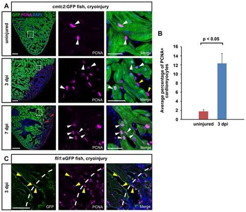
Activation of cardiomyocyte proliferation in response to cryoinjury. (A) Mature cardiomyocytes located close to the lesion in the uninjured myocardium proliferate at 3 and 7 dpi. Uninjured and cryolesioned hearts (3 dpi and 7 dpi) of cmlc2:GFP transgenic fish were stained for GFP and PCNA. Nuclei are stained with Dapi. Proliferating cardiomyocytes are indicated by white arrowheads. Proliferating endocardial cells are indicated by yellow arrowheads (3 dpi). Note individual GFP+ cardiomyocytes inside the lesioned area at 7 dpi (red arrowheads). Scale bars are 100 μm in the overview and 25 μm in the close ups. n = 3 hearts (9 sections) at all conditions. (B) Quantification of the percentage of PCNA+ cardiomyocytes in uninjured versus cryolesioned hearts at 3 dpi. CMs were quantified in the entire ventricle. Error bars = s.e.m. n = 3 hearts (7 sections for 3 dpi, 5260 CMs counted; 9 sections for uninjured control, 7475 CMs counted), Student′s t-test was employed for statistical analysis, with p<0.05. (C) Endocardial cells located adjacent to the lesion proliferate at 3 dpi. Cryolesioned hearts (3 dpi) of fli1:eGFP transgenic fish were stained for GFP and PCNA. Nuclei are stained with Dapi. Proliferative endocardial cells are indicated by yellow arrowheads. Wound edge is indicated by a white dashed line. Scale bars are 25 μm. |