
Fig. 1
- ID
- ZDB-FIG-110425-2
- Publication
- Herpin et al., 2007 - Specification of primordial germ cells in medaka (Oryzias latipes)
- Other Figures
- All Figure Page
- Back to All Figure Page
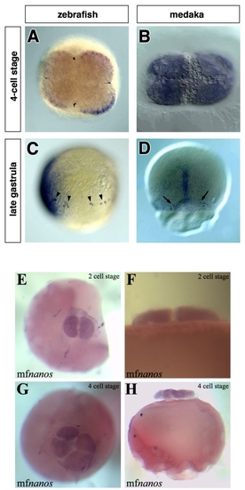
The early expression patterns of zebrafish-vasa, medaka-olvas and medaka-nanos1 mRNAs. In zebrafish, the vasa transcript is enriched at the marginal positions of the first two cleavage planes (A), while at a similar stage olvas mRNA is uniformly distributed throughout the cytoplasm of all blastomers (B). Throughout development in zebrafish, the vasa mRNA is expressed exclusively in the PGCs that can be found in random dorsoventral positions during blastula and gastrula stages (arrowheads in C). In contrast, at the first time point when medaka PGCs can be observed (stage 16), they are found on both sides of the embryonic shield on the dorsal side of the embryo (arrows in D). All images shown are whole-mount in situ hybridizations. The probes used are: vasa (A), vasa and chordin (C), olvas (B, D) and nanos1 (E to H). A, B, E and H are animal view, C is a lateral view with the dorsal aspect (labeled with chordin) to the left and D is a dorsal view, F and H are lateral views. |