
Fig. 6
- ID
- ZDB-FIG-110407-41
- Publication
- Carvalho et al., 2011 - A high-throughput screen for tuberculosis progression
- Other Figures
- All Figure Page
- Back to All Figure Page
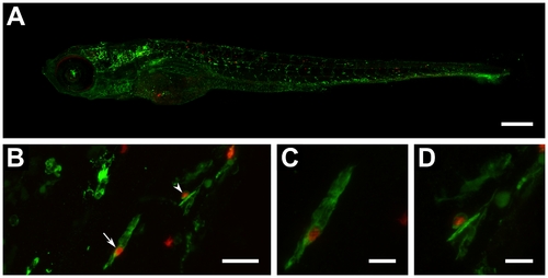
Automatic yolk sac injection of M. tuberculosis and effect of treatment on infected larvae. (A) Confocal z-stack (8×2 stitching) of a 6 day-old whole larva (fli1-egfp with gfp-labelled vasculature) showing spread of bacteria (red) throughout the body (scale bar: 250 μm). (B) Confocal z-stack of red-fluorescent bacteria co-localizing with green-fluorescent leukocytes detected by L-plastin immunostaining (scale bar: 25 μm). (C) Close-up (digital zoom: 4.2) of bacteria-containing leukocyte depicted in B by straight arrow (scale bar: 10 μm). (D) Close-up (digital zoom: 4.3) of bacteria-containing leukocyte depicted in B by arrowhead (scale bar: 10 μm). |