
Fig. S1
- ID
- ZDB-FIG-110218-7
- Publication
- Barrios et al., 2003 - Eph/Ephrin signaling regulates the mesenchymal-to-epithelial transition of the paraxial mesoderm during somite morphogenesis
- Other Figures
- All Figure Page
- Back to All Figure Page
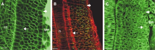
Cells within the Core of the PSM Are Mesenchymal, and Epithelialization of Somite Boundary Cells Occurs Concomitantly with Boundary Formation (A–C) Dorsal views of the PSM of wild-type embryos; anterior is oriented toward the top. Embryos have been stained with (A) Bodipy ceramide or (B) phalloidin (red) or (C) Bodipy 505-515. In (B), the embryo has been immunostained for γ-tubulin (green) to show centrosomes. The arrows point to presomitic cells that have epithelial morphology adjacent to the (A) neural tube, the (B) notochord, and the (C) lateral plate mesoderm. Other cells in the PSM have mesenchymal morphology. The arrowheads indicate the most recently formed or forming intersomitic boundary in each panel. Cells on either side of this boundary only begin to acquire epithelial morphology concomitant with boundary formation. Full maturation of epithelial morphology (see Figures 1A–1D in the main text) occurs after boundary furrow formation. lpm, lateral plate mesoderm; nt, neural tube; n, notochord. The scale bar represents 15 μm. |