FIGURE

Fig. S1

ID
ZDB-FIG-110214-72
Publication
Ohata et al., 2011 - Dual Roles of Notch in Regulation of Apically Restricted Mitosis and Apicobasal Polarity of Neuroepithelial Cells
Other Figures
All Figure Page
Back to All Figure Page
Fig. S1

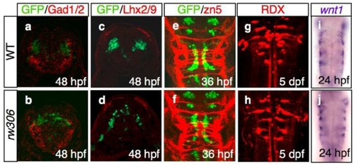
(a-d) The location of the Glutamate decarboxylase 1 and 2 (Gad1/2)-immunoreactive neurons (a,b; red), as well as the LIM homeodomain proteins, Lhx2 and Lhx9 (Lhx2/9)-immunoreactive neurons (c,d; red), which are segmentally positioned in the hindbrain (Ando et al., 2005; Sassa et al., 2007) were examined in the WT (a,c) and rw306 mutant (b,d) embryos. Cross-sectional views, dorsal to the top. The motor neurons are labeled green. Note that the locations of the Gad1/2- and Lhx2/9-immunoreactive neurons were almost normal in the rw306 embryos in the present study.
(e,f) Segmentally repeated commissural neurons and their axons in the zebrafish hindbrain were visualized using the zn-5 antibody (Trevarrow et al., 1990) (red) in the WT (e) and rw306 mutant (f) embryos at 36 hpf. Dorsal view, rostral to the top. The motor neurons are labeled green. Note that the formation of the zn-5-immunoreactive axons appeared to be normal in the rw306 mutant embryos
(g,h) Retrograde labeling of reticulospinal neurons by injection of a tracer dye (rhodamine-dextran, RDX; red) into the spinal cord of the WT (g) and rw306 (h) embryos at 5 days post-fertilization (dpf). Ventral view, rostral to the top. Note that the anterior-posterior patterning of the reticulospinal neurons in the rw306 embryos was identical to that in the WT embryos.
(i,j) In situ hybridization of the hindbrain segmentation marker wnt1 mRNA (purple) in the WT (i) and rw306 (j) embryos at 24 hpf. Dorsal view; rostral to the top. Note that the expression pattern of wnt1 was unaffected in the rw306 embryos.

Expression Data

Expression Detail
Antibody Labeling
Phenotype Data

Phenotype Detail
Acknowledgments
This image is the copyrighted work of the attributed author or publisher, and ZFIN has permission only to display this image to its users. Additional permissions should be obtained from the applicable author or publisher of the image. Full text @ Neuron