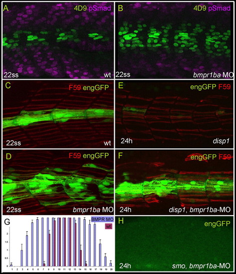
Inhibition of Smad activity de-represses engrailed expression. (A,B) Parasagittal optical sections of 22 ss embryos showing distribution of Eng (4D9) and pSmad in wild type (A) and bmpr1ba morphants (B). Somites shown are at the level of the yolk sac. (C,D,G) Attenuation of BMPR1ba causes ectopic expression of eng2a. The histogram in G compares eGFP expression in Tg(eng2a:eGFP)i233 embryos injected with bmpr1ba morpholino, with that in non-injected controls; bars represent the frequency of eGFP expressing slow fibres at different dorsoventral positions. Positions 8-13 represents the horizontal myoseptum. Morphants display a wide expansion of the eGFP domain. Data based on three consecutive somites at the end of yolk extension in 22 ss embryos. x axis, dorsoventral position; y axis, number of eGFP+ fibres. Error bars indicate s.d. Representative examples are shown in C,D. (E) Parasagittal optical sections of a 24 hour Tg(eng2a:eGFP)i233;disp1tf18b mutant embryo displaying reduced slow fibres (F59; red) and near absence of eng2a:eGFP. (F) Similarly staged Tg(eng2a:eGFP)i233; disp1tf18b mutant embryo that was injected with bmpr1ba morpholino at the one- to two-cell stage, showing robust rescue of eGFP expression. (H) Parasagittal optical sections of a 24 hour Tg(eng2a:eGFP)i233; smob641 mutant embryo injected with bmpr1ba morpholino at one- to two-cell stage, showing complete absence of eng2a:eGFP expression. Inhibition of Smad activity de-represses engrailed expression. (A,B) Parasagittal optical sections of 22 ss embryos showing distribution of Eng (4D9) and pSmad in wild type (A) and bmpr1ba morphants (B). Somites shown are at the level of the yolk sac. (C,D,G) Attenuation of BMPR1ba causes ectopic expression of eng2a. The histogram in G compares eGFP expression in Tg(eng2a:eGFP)i233 embryos injected with bmpr1ba morpholino, with that in non-injected controls; bars represent the frequency of eGFP expressing slow fibres at different dorsoventral positions. Positions 8-13 represents the horizontal myoseptum. Morphants display a wide expansion of the eGFP domain. Data based on three consecutive somites at the end of yolk extension in 22 ss embryos. x axis, dorsoventral position; y axis, number of eGFP+ fibres. Error bars indicate s.d. Representative examples are shown in C,D. (E) Parasagittal optical sections of a 24 hour Tg(eng2a:eGFP)i233;disp1tf18b mutant embryo displaying reduced slow fibres (F59; red) and near absence of eng2a:eGFP. (F) Similarly staged Tg(eng2a:eGFP)i233; disp1tf18b mutant embryo that was injected with bmpr1ba morpholino at the one- to two-cell stage, showing robust rescue of eGFP expression. (H) Parasagittal optical sections of a 24 hour Tg(eng2a:eGFP)i233; smob641 mutant embryo injected with bmpr1ba morpholino at one- to two-cell stage, showing complete absence of eng2a:eGFP expression.
|
