
Fig. 1
- ID
- ZDB-FIG-110214-52
- Publication
- Vonica et al., 2011 - APOBEC2, a selective inhibitor of TGFβ signaling, regulates left–right axis specification during early embryogenesis
- Other Figures
- All Figure Page
- Back to All Figure Page
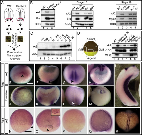
APOBEC2 is a target of TGFβ signaling coexpressed with derrière in Xenopus embryos. (A) Strategy for identification of genes regulated by derrière. Posterior dorsal fragments from wild-type and derrière -MO (Der-MO)-injected embryos were isolated at stage 18. (B) TGFβ signaling and xA2 expression. Overexpression of a dominant negative type 1 receptor (DN ALK4) reduces expression of marginal xA2 in stage 10 whole embryos (left panel). Overexpressed Xnr1 (30 pg) and derrière (100 pg) RNA induce xA2 expression in stage 10 (central panel) and 18 (right side panel) animal caps. RT-PCR for xA2, MyoD, and Brachyury as markers of mesoderm induction, and ODC as loading control. (C) Timing of xA2 expression. RT-PCR of embryos collected at the indicated developmental stages. (D) Spatial expression of xA2 in stage 10 embryos. RT-PCR of embryonic explants (VMZ: ventral marginal zone; DMZ: dorsal marginal zone). (E–M) Comparative expression of xA2 and derrière. In situ hybridization for xA2 (E–I) and derrière (J–M) expression. (E, J) Stage 10 vegetal–dorsal views, arrowhead indicates the forming dorsal lip; (F, K) stage 11 dorso-ventral sections (dorsal to the right). Arrowheads indicate recently involuted mesoderm; (G, L) dorsal views (anterior up). Arrowheads indicate the blastopore; (H, M) stage 16 transversal sections, posterior fragments (dorsal is up). (I) Stage 32 lateral view. Overlap between xA2 and derrière occurs at stage 10 (dorsal marginal), stage 11 (involuted mesoderm), and stage 16 (paraxial mesoderm). (N–R) Expression of zebrafish A2 (zA2). (N, O, P) Seventy-five percent epiboly, in (N) lateral view (dorsal to the right) and (O) dorsal views. The arrowheads in panels N and O indicate the shield. (Q–R) Fourteen somite stage embryo. The inset in panel O (twofold magnification) shows shield cells with nuclear stain. (Q) Lateral view, anterior to the left, (R) dorsal view (anterior up). (P) Embryo stained with the sense probe as negative control. The scale bars in panel E indicates 0.3 mm, and in panel N, 0.1 mm. |
| Gene: | |
|---|---|
| Fish: | |
| Anatomical Terms: | |
| Stage Range: | 75%-epiboly to 14-19 somites |
Reprinted from Developmental Biology, 350(1), Vonica, A., Rosa, A., Arduini, B., and Brivanlou, A.H., APOBEC2, a selective inhibitor of TGFβ signaling, regulates left–right axis specification during early embryogenesis, 13-23, Copyright (2011) with permission from Elsevier. Full text @ Dev. Biol.