
Fig. S7
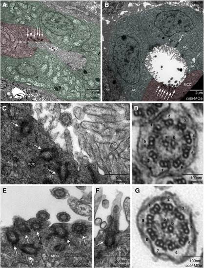
Structure and localization of basal bodies and cilia appear unaffected in cobl morphants. Transmission electron micrographs of sections through the proximal straight tubules of the pronephros in controls and morphants. (A–B) Morphant tubules appear dilated. Multi-ciliated cells (MCC) are false-colored red. Absorbing cells (AC) are false-colored green. Basal bodies appear localized to the apical surface in multi-ciliated cells (A–D) and absorbing cells (E). Cross sections through the 9 + 2 ciliary axoneme (F–G) reveal no structural difference between control and morphant cilia. Arrows point to basal bodies. Outer microtubule doublets are numbered 1–9, inner core filaments labeled a/b. |
Reprinted from Developmental Biology, 350(1), Ravanelli, A.M., and Klingensmith, J., The Actin Nucleator Cordon-bleu is Required for Development of Motile Cilia in Zebrafish, 101-111, Copyright (2011) with permission from Elsevier. Full text @ Dev. Biol.