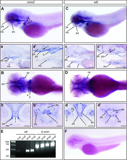
Expression pattern of Runx2b and VDR in Danio rerio. An in situ hybridization of a 72 hpf zebrafish embryo hybridized with a Runx2b antisense probe is shown in a lateral (A) and ventral (B) view. The images in a and a′ are sagittal sections of the specimen shown in A. The images in b and b′ are transversal sections of the specimen shown in B. An in situhybridization of a 72 hpf zebrafish embryo hybridized with a VDR antisense probe is shown in a lateral (C) and ventral (D) view. The images in c and c′ are sagittal sections of the specimen shown in C. The images in d and d′ are transversal sections of the specimen shown in D. (E) RT-PCR performed on cDNA from zebrafish embryos at 0, 24 hpf, 48 hpf and larvae at 72 hpf with primers specific for VDR and β-actin. The molecular weight standard is shown on the left. (F) Lateral view of a 72 hpf zebrafish larvae hybridized with a VDR sense probe. Abbreviations: c, crystalline; cl, cleithrum; de, diencephalon; ipl, inner plexiform layer; mc, meckel cartilage; op, operculum; pq, palate quadrate; ps, parasphenoid; q, quadrate; t, trabecula; v, ventricle; 4 and 5, the IV and V branchial cartilage territories, respectively. The scale bar in a, a′, b, b′, c, c′, d and d′ represents 50 micrometers.
|
