
Fig. 3
- ID
- ZDB-FIG-101111-43
- Publication
- Prykhozhij, 2010 - In the Absence of Sonic Hedgehog, p53 Induces Apoptosis and Inhibits Retinal Cell Proliferation, Cell-Cycle Exit and Differentiation in Zebrafish
- Other Figures
- All Figure Page
- Back to All Figure Page
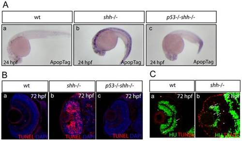
Apoptosis was assayed in whole-mount embryos using ApopTag (A, a-c) and on retinal sections using TUNEL labelling (B, a-c). The sections are oriented with their anterior side to the top. Elevated apoptosis in shh-/- mutant embryos compared to wild-type embryos was suppressed by p53 loss in the developing nervous system (A, a-c) and in the retina (B, a-c). (A) Whole-mount ApopTag staining at 24 hpf of wild-type, shh-/- and p53-/-shh-/- embryos (a-c). (B) TUNEL/DAPI staining of retinal sections of wild-type, shh-/- and p53-/-shh-/- embryos at 72 hpf (a-c). (C) Wild-type (a) and shh-/- mutant (b) retinal cryosections from 72 hpf embryos stained with anti-HU antibodies to detect retinal neurons and TUNEL to label apoptotic cells. Cells positive for both HU and TUNEL are indicated with arrows and cells positive only for TUNEL are labelled with arrowheads. Images were obtained from retinal sections of 10 embryos and representative overlay images are shown. |