
FIGURE
Fig. 2
- ID
- ZDB-FIG-101104-3
- Publication
- Raphael et al., 2010 - Schwann cells reposition a peripheral nerve to isolate it from postembryonic remodeling of its targets
- Other Figures
- All Figure Page
- Back to All Figure Page
Fig. 2
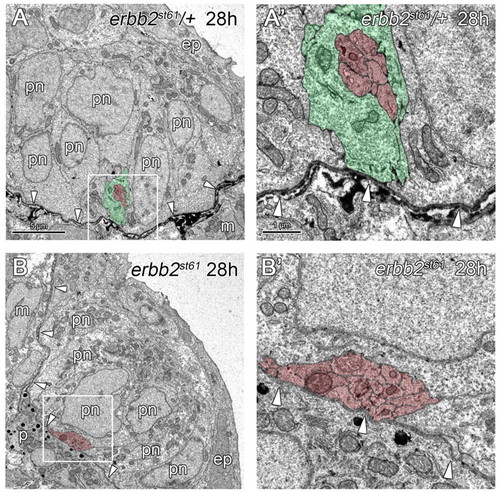
PLLn outgrowth is normal in Schwann cell-deficient mutants. (A,B) At 28 hpf, axons and Schwann cells of heterozygous siblings (A) and axons of erbb2st61 mutants (B) are located within the epidermis (ep) (n=3 in A and n=6 in B), and are associated with pre-neuromast cells (pn). (A′,B′) Higher magnification of the boxed regions in A,B, respectively. Arrowheads indicate the location of the epidermal basement membrane. Scale bars: 5 μm in A,B; 1 μm in A′,B′. Schwann cells are pseudocolored green and axons in red. Abbreviations: ep, epidermis; m, muscle; pn, pre-neuromast cell. |
Expression Data
Expression Detail
Antibody Labeling
Phenotype Data
Phenotype Detail
Acknowledgments
This image is the copyrighted work of the attributed author or publisher, and
ZFIN has permission only to display this image to its users.
Additional permissions should be obtained from the applicable author or publisher of the image.
Full text @ Development