
Fig. 4
- ID
- ZDB-FIG-101025-12
- Publication
- Bernick et al., 2010 - Knockdown and overexpression of Unc-45b result in defective myofibril organization in skeletal muscles of zebrafish embryos
- Other Figures
- All Figure Page
- Back to All Figure Page
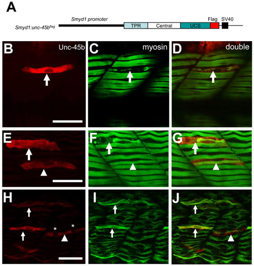
Overexpression of Unc-45b resulted in defective thick filament organization. A. The DNA construct expressing a flag-tagged Unc-45b under the control of smyd1 promoter was injected into zebrafish embryos. The effect on thick filament organization was analyzed by double antibody staining at 30 hpf. B-D. Double staining with anti-flag (red) and anti-myosin (F59, green) antibodies shows the expression of flag-tagged Unc-45b (B) in a single fiber and its effect on myosin thick filament organization (C). D represents the merged image of B and C showing that the myofibril defect is restricted to the myofiber expressing the flag-tagged Unc-45b. Fifty six myofibers from 40 embryos were analyzed. 70% of the myofiber showed the disorganized thick filaments. E-G. Double staining shows the expression of flag-tagged Unc-45b in two myofiber with different phenotypes on myosin thick filament organization. One myofiber exhibited disorganized thick filament (Arrow). In contrast, another myofiber exhibited normal thick filament organization (arrow head). However, this fiber appeared to be skinnier compared with its neighbors without the ectopic Unc-45b expression. H-J. Double staining shows the rescue of thick filament organization in Unc45b knockdown zebrafish embryos co-injected Unc45b-MO with the smyd1:unc45bflag DNA construct. Myofibers expressing the flag-tagged Unc45b (H) exhibited normal thick filament organization (I, J). The fast fiber with two nuclei (*) is indicated by the arrow head. F59 does not label myosin expressed in fast muscles. Thirty five slow myofibers from 29 embryos were analyzed. 100% of the slow myofiber expressing the flag-tagged Unc45b (H) exhibited normal thick filament organization. Scale bars = 15 μm. |