Fig. S1
- ID
- ZDB-FIG-100616-5
- Publication
- McGrail et al., 2010 - Expression of the zebrafish CD133/prominin1 genes in cellular proliferation zones in the embryonic central nervous system and sensory organs
- Other Figures
- All Figure Page
- Back to All Figure Page
|
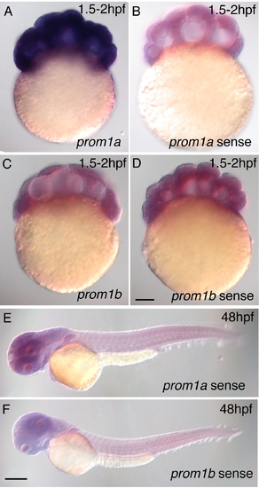
Control experiments to demonstrate specificity of the prom1a and prom1b antisense probes used for in situ hybridization. Specific labelling using antisense probes (A, C) versus background signal in embryos hybridized with sense probes (B, D-F). Intense labelling with the prom1a probe in the 2hpf embryo (A) is consistent with RT-PCR demonstrating high levels of maternal message within the 1hpf embryo. In contrast, the absence of specific labeling by prom1b in the 2hpf embryo (C) is consistent with RT-PCR showing barely detectable levels of maternal message in the 1hpf embryo. Figure 1B, lane 1, compare prom1a and prom1b. Scale bars = 100 μm. |

