
FIGURE
Fig. S1
Fig. S1
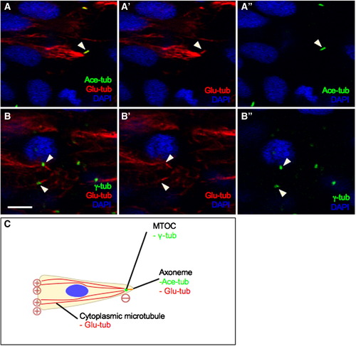
The centriole templates early ciliary axoneme assembly. High magnifications views of cross sections through the hindbrain region of embryos at the neural keel stage, immunolabeled with (A–A″) anti-Ace-tub (ciliary axoneme, in green) and anti-Glu-tub (cytoplasmic MT and ciliary axoneme, in red) or with (B–B″) anti-γ-tub (centrosome, in green) and anti-Glu-tub. Nuclei are labeled with DAPI, in blue. (C) Illustration of cellular organelles visualized using different antibodies. Symbols: arrowheads indicate axonemes; arrows show the centrosome. Scale bar: 5 μm. |
Expression Data
Expression Detail
Antibody Labeling
Phenotype Data
Phenotype Detail
Acknowledgments
This image is the copyrighted work of the attributed author or publisher, and
ZFIN has permission only to display this image to its users.
Additional permissions should be obtained from the applicable author or publisher of the image.
Reprinted from Developmental Biology, 341(2), Hong, E., Jayachandran, P., and Brewster, R., The polarity protein Pard3 is required for centrosome positioning during neurulation, 335-345, Copyright (2010) with permission from Elsevier. Full text @ Dev. Biol.