
Fig. 7
- ID
- ZDB-FIG-100322-17
- Publication
- Kimmel et al., 2010 - Modes of developmental outgrowth and shaping of a craniofacial bone in zebrafish
- Other Figures
- All Figure Page
- Back to All Figure Page
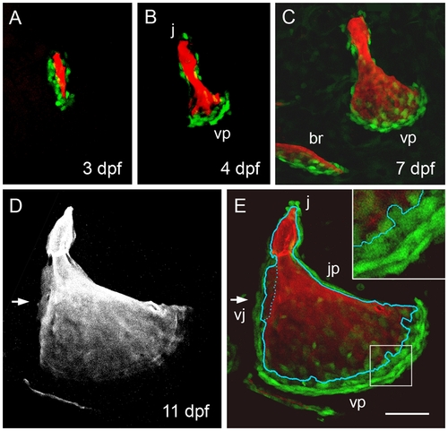
Osteoblast arrangements change dynamically during opercle morphogenesis. Confocal imaging of live preparations. (A-C) Two-color merged images showing Alizarin Red A labeling of the matrix and osx:eGFP labeling of the bone forming cells. (A) At 3 dpf osteoblasts line up along the developing bony spur. (B, C) At 4 and 7 dpf a new arrangement is present with cells especially concentrated along the rapidly outgrowing vp edge. The newly forming posterior branchiostegal ray (br) is included in C. (D) Red channel, and (E) merge at 11 dpf. Arrows indicate the vj veil. In E the outline of the Alizarin Red labeled bone (from D) is superimposed. Very flattened and compact looking osx:eGFP-expressing cells line the very slowly growing jp edge and are present in outer rows of the very rapidly growing vp edge. Small round cells are present at the j apex spur. Larger diffusely labeled cells are present along the vj veil and in the innermost row along the vp edge, where the cells immediately contact new mineralized matrix (a portion of this edge is enlarged in the inset). Scale bar: 50 μm. |
| Gene: | |
|---|---|
| Fish: | |
| Anatomical Term: | |
| Stage Range: | Protruding-mouth to Days 7-13 |