
Fig. 3
- ID
- ZDB-FIG-100223-25
- Publication
- Camarata et al., 2010 - Pdlim7 (LMP4) regulation of Tbx5 specifies zebrafish heart atrio-ventricular boundary and valve formation
- Other Figures
- All Figure Page
- Back to All Figure Page
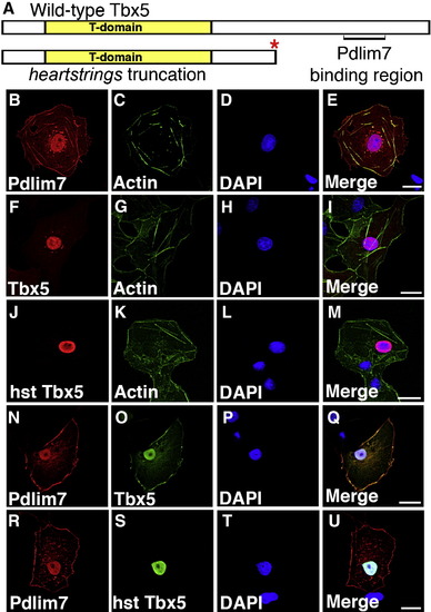
Pdlim7 colocalizes with full-length but not the hearstrings Tbx5. (A) Schematic of wild-type and heartstrings (hst) Tbx5 proteins. Pdlim7 binding region is denoted in wild-type Tbx5. Red asterisk indicates the truncation site of the hst encoded protein. (B–E) Single transfection of myc-Pdlim7 in COS-7 cells. Cells stained with anti-myc antibodies (B), Alexa-488 phalloidin to stain filamentous actin (C), and DAPI nuclear stain (D). (E) Merged image of B–D. (F–I) Single transfection of full-length HA-Tbx5. Cells stained with anti-HA antibodies (F), Alexa-488 phalloidin (G), and DAPI (H). (I) Merged image of F–H. (J–M) Individual transfection of truncated HA-Tbx5 resembling the encoded hst allele. Cells stained for Tbx5 (J), actin (K), and nucleus (L). (M) Merged image of J–L. (N–Q) Cotransfected COS-7 cells stained for Pdlim7 (N), full-length Tbx5 (O), and nucleus (P). (Q) Merged image of N-P. (R-U) Cotransfected cells stained for Pdlim7 (R), truncated Tbx5 (S), and nucleus (T). (U) Merged image of R-T. Scale bar, 20 μm. |
Reprinted from Developmental Biology, 337(2), Camarata, T., Krcmery, J., Snyder, D., Park, S., Topczewski, J., and Simon, H.G., Pdlim7 (LMP4) regulation of Tbx5 specifies zebrafish heart atrio-ventricular boundary and valve formation, 233-245, Copyright (2010) with permission from Elsevier. Full text @ Dev. Biol.