
Fig. S3
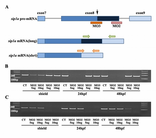
sip1a splice variant targeting morpholinos efficiently eliminate short and long forms. A: Diagram of the alternative splicing within the 3′ region of the sip1a pre-mRNA. Alternative splicing of exon 8 eliminates one zinc finger (dark blue box), which is present in the longer form. Sip1a MO3 (orange bar) targeted to alternative splice site in exon 8 (denoted by black arrow) blocks the alternating splicing event and eliminates production of the short form. Sip1a MO2 (pink bar) blocks the pre-mRNA splicing event needed to generate the long form by targeting the splice site at the 3′ end of exon 8. B,C: Reverse transcriptase-polymerase chain reaction (RT-PCR) of mRNA from staged sip1a splice MO2 and MO3 injected embryos with sip1a full length specific primers (B, green arrows in A) and sip1a short form specific primers (C, orange arrows in A ). B,C: sip1 splice MO2 efficiently altered splicing so that the full length message is eliminated and only the shorter form was produced (B) while sip1 splice MO3 blocked production of the shorter form (C). |