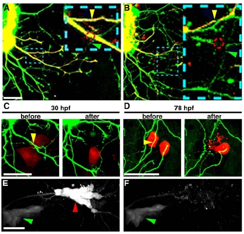
Two-Photon Excitation Precisely Severs Axons and Causes Limited Tissue Damage
(A and B) Confocal images taken before (A) and 1 hr after (B) two-photon axotomy at 30 hpf. All trigeminal neurons were labeled with GFP, and a variegated population labeled with RFP. Dashed blue box indicates region magnified in inset. Dashed red circle indicates site of axotomy. Arrowheads point to a branch of the injured axon (yellow) and a branch of a neighboring uninjured axon (green) that remained intact even though they were in close proximity (<8 μm away) to the site of axotomy (axotomized branch in B has lost red fluorescence due to bleaching). In more than one hundred axotomies, we never observed damage to axon branches that were not specifically targeted.
(C and D) Confocal images taken before and 30 min after 30 hpf (C) or 78 hpf (D) two-photon axotomy. Variegated population of trigeminal neurons labeled with GFP, keratinocytes labeled with mCherry. Yellow arrow indicates site of axotomy. A single skin cell directly above or below the site of axotomy died after both 30 and 78 hpf axotomy (7/7 axotomies at 30 hpf and 6/6 axotomies at 78 hpf).
(E and F) Two-photon images taken before (E) and 1 hr after (F) two-photon ablation of an entire trigeminal ganglion at 30 hpf. All trigeminal neurons as well as other cranial ganglia were labeled with GFP. Red arrowhead indicates ablated ganglion. Green arrowhead indicates a neighboring cranial ganglion that remained intact.
Scale bar represents 50 μm in all panels.
|
