
Fig. 6
- ID
- ZDB-FIG-091221-32
- Publication
- Song et al., 2009 - Mechanisms Underlying Metabolic and Neural Defects in Zebrafish and Human Multiple Acyl-CoA Dehydrogenase Deficiency (MADD)
- Other Figures
- All Figure Page
- Back to All Figure Page
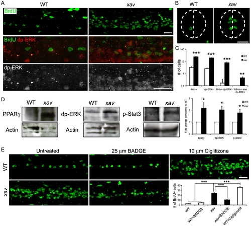
xav mutants exhibit increased neural cell proliferation as a result of increased glycolysis, due to perturbation of the PPARG-ERK pathway. A. Whole mount embryos labeled with BrdU to mark cells undergoing proliferation. The number of BrdU+ cells was significantly increased in the nervous system, especially in the spinal cord, in xav mutants compared to WT embryos at ∼56–60 hpf. Increased dp-ERK+ cells and BrdU+/dp-ERK+ double labeled cells in the spinal cord in xav mutants compared to WT embryos at ∼56–60 hpf. B. In spinal cord cross-sections from xav mutants and WT embryos at ∼56–60 hpf, BrdU+ cells are distributed peri-ventricularly, suggesting that they are likely to be neural progenitor cells. Dashed line outlines the spinal cord and indicates the midline. Scale bar = 20 μm. C. Quantification of BrdU+ and dp-ERK+ cells at ∼56–60 hpf. Per spinal cord hemisegment: BrdU+ cells WT 3.2±0.4, xav 24±1.4; dp-ERK+ cells WT 2.6±0.3, xav 19.4±0.8; BrdU+/dp-ERK+ WT 0.2±0.2, xav 7.8±1.2. Percent of BrdU+ cells that are also dp-ERK+: WT 3.6%±3.6%, xav 45%±8% (N = 4-19 embryos, >2 carrier pairs; Student′s t test, *** p<0.001). D. Western blot analyses of pparγ, dp-ERK and phospho-STAT3 expression showed dramatic increase in xav compared to WT embryos at ∼56–60 hpf (N = 3 replicates, 30 embryos each; Student′s t test, * p<0.05). E. BrdU labeling of proliferating cells in whole mounts of spinal cord of WT embryos, xav mutants, WT treated with 25 μm BADGE, xav mutants treated with 25 μm BADGE and WT treated with 10 μm Ciglitizone at ∼56–60 hpf. Embryos were raised in BADGE or Ciglitizone from 24 to 60 hpf. Per spinal hemisegment: BrdU+ cells WT 3.2±0.4, WT + BADGE 4.5±0.3, xav 24±1.4, xav + BAGDE 10.6±0.9, WT + Ciglitizone 36±2.5 (N = 6–19 embryos, >3 carrier pairs; one-way ANOVA, followed by Bonferroni′s multiple comparison test, *** p<0.001). |
| Fish: | |
|---|---|
| Observed In: | |
| Stage: | Long-pec |