FIGURE

Fig. 8

ID
ZDB-FIG-091116-2
Publication
Schier et al., 1996 - Mutations affecting the development of the embryonic zebrafish brain
Other Figures
All Figure Page
Back to All Figure Page
Fig. 8

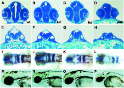
Phenotypic analysis of mutations affecting ventricle enlargement. (A,E,I,M) Wild type; (B,F,J,N) silent heart (sih)b109; (C,G,K,O) fullbrain (ful)m133; (D,H,L,P) snakehead (snk)m273. (A,B,C,D) Transverse sections through eye and lens at 28 hpf. (E,F,G,H) Transverse section through anterior ear and otolith at 28 hpf. Note the different degrees of ventricle (white arrowheads) reduction in sih, ful and snk. (I,J,K,L) Expression of rtk1 in rhombomeres 1,3 and 5 at 31 hpf; dorsal view. (M,N,O,P) Wild-type and mutant embryos at 53 hpf. Note the differences in reduction in the size of the brain and the onset of degeneration in sih, ful and snk.

Expression Data
Gene:
Fish:
Anatomical Terms:
Stage: Prim-15

Expression Detail
Antibody Labeling
Phenotype Data
Fish:
Observed In:
Stage Range: Prim-5 to Long-pec

Phenotype Detail
Acknowledgments
This image is the copyrighted work of the attributed author or publisher, and ZFIN has permission only to display this image to its users. Additional permissions should be obtained from the applicable author or publisher of the image. Full text @ Development