
Fig. 3
- ID
- ZDB-FIG-090710-5
- Publication
- Roberts et al., 2009 - Apical polarity protein PrkCi is necessary for maintenance of spinal cord precursors in zebrafish
- Other Figures
- All Figure Page
- Back to All Figure Page
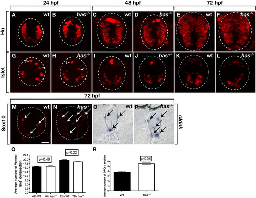
Loss of protein kinase C, iota (PrkCi) function produces excess oligodendrocyte progenitor cells (OPCs). All images are of transverse sections through trunk spinal cord, dorsal up. Outlined circle marks the perimeter of the spinal cord. A-F: The number and distribution of neurons, marked by anti-Hu labeling, are similar in wild-type and has-/- embryos at 24, 48, and 72 hours postfertilization (hpf). G-L: Isl+ motor neurons (brackets), interneurons (solid arrowheads), and Rohon-Beard sensory neurons (open arrowheads) are similar in wild-type and has-/- embryos and larvae through 72 hpf. M,N: Anti-Sox10 labeling reveals excess OPCs in 72 hpf has-/- larva (N) compared with wild-type (M). O,P: cldnk RNA expression marking differentiating oligodendrocytes. O: In wild-type, cldnk+ cells (arrows) are at the pial surface in dorsal and ventral spinal cord. P: has-/- larvae have excess cldnk+ cells and some occupy ectopic positions in medial spinal cord. Q: Quantification of spinal cord Isl+ motor neurons in the spinal cord between wild-type and has-/- embryos at 48 and 72 hpf. R: Quantification of spinal cord Sox10+ OPCs. Error bars represent SEM. Statistical significance was determined using Student′s t-test. Scale bar = 20 μM. |