
Fig. 6
- ID
- ZDB-FIG-090710-13
- Publication
- Martin et al., 2009 - Analysis of heart valve development in larval zebrafish
- Other Figures
- All Figure Page
- Back to All Figure Page
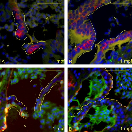
Larger larvae have more pronounced maturation of the valves at 28 days postfertilization (dpf). A-D: Five-micrometer paraffin sections of flk1:GFP (GFL, green fluorescent protein) transgene-carrying zebrafish larvae were imaged to identify the structural appearance of the atrioventricular (AV) boundary and intra-luminal structure. Atria (A) and ventricles (V) are labeled (yellow letters), and yellow lines outline the extent of the AV structure (cushion and/or valve). A,B: Sections were immunostained with DAPI (4′,6-diamidine-2-phenylidole-dihydrochloride; blue), anti-Versican (red) and anti-Collagen I (green). Whereas the smaller larva (A) has started deposition of Versican in the leaflet, the larger larva (B) has thickened considerably more, with increased deposition of Versican and Collagen I. C,D: From separate larvae than shown in A,B, sections were stained with DAPI (blue), anti-GFP (red), and anti-Collagen II (green). Again, the larger larva (D) has significantly more deposition of collagen than the smaller larva (C), despite being at the same age. Scale bar = 20 μm. |