
Fig. S1
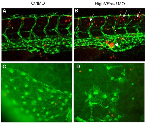
Endothelial cell death is not increased in VE-cadherin endothelial cells. (A–D) TUNEL assay of 48 hpf embryos in a fli1:EGFP background allows the colocalization analysis of apoptotic cells (in red) and endothelial cells (in green). (A–B) Confocal images of the trunk region at the level of the urogenital opening are shown. VE-cadherin severe morphants (B) exhibit increased apoptosis in the blood island (arrowhead) where the blood cells and hematopoietic precursors (some of them fli1:EGFP positive) accumulate in the absence of circulation (arrowhead). In addition, increase in the number of apoptotic cells is observed in the tissues surrounding the ISVs and DLAV (arrows). Few vascular endothelial cells (asterisks) show increase in TUNEL staining. (C–D) Fluorescence microscopy images of the endothelial cells in the sinus venosus. |