
Fig. 7
- ID
- ZDB-FIG-090408-97
- Publication
- Shima et al., 2009 - The characterization of a zebrafish mid-hindbrain mutant, mid-hindbrain gone (mgo)
- Other Figures
- All Figure Page
- Back to All Figure Page
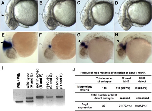
Microinjection of pax2a mRNA rescued eng3 expression. pax2a mRNA (50 pg) was injected into 1-cell-stage embryos derived from heterozygous Wik/AB* mgo incross. Embryonic genotypes and marker gene are indicated at the bottom and top, respectively. A-H: Lateral views of the MHB at 24 hpf embryos of wild type sibling (A), mgo (B), and mgo injected with pax2a mRNA (C,D). In situ hybridization with eng3 (E-H). Wild-type embryos had a distinct MHB structure and eng3 mRNA was expressed strongly at MHB region (A,E). In the mgo mutant, the MHB structure was absent and eng3 expression was limited to the ventral region (B,F). pax2a mRNA injected into a clutch of embryos derived from an mgo incross showed 20.3% with the MHB phenotype. However, eng3 expression was expanded in both the ventral and dorsal region (C,D,G,H). I: Embryos genotyped by PCR with a pax2a intron 3 marker. Wild-type, Wik/Wik background, yields lower band. Heterozygote Wik/AB* background yields two bands, and homozygous mutants showed an upper single band. J: Summary of rescue experiment. |