
Fig. 1
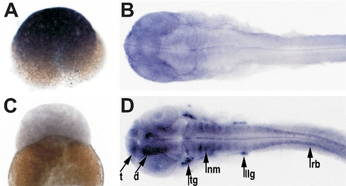
Differential Expression of PrP Genes in Zebrafish Embryos The developmental expression patterns of PrP-1 and -2 were examined by whole mount in situ RNA hybridization using gene-specific probes. At mid-blastula stages (2.5 hpf, [A and C]), PrP-1 is ubiquitously transcribed at high levels and PrP-2 is not detectable. At pharyngula stages (30 hpf, [B and D]), low levels of PrP-1 transcripts appear restricted to the forebrain and eyes, while PrP-2 becomes strongly transcribed in defined neural structures. (A and C) show lateral views; (B and D) show dorsal views. d, diencephalon; llg, lateral line ganglion; nm, neuromeres; rb, Rohon-Beard sensory neurons; t, telencephalon; tg, trigeminal ganglion. |
| Genes: | |
|---|---|
| Fish: | |
| Anatomical Terms: | |
| Stage Range: | 256-cell to Prim-15 |