
Fig. 1
- ID
- ZDB-FIG-090401-39
- Publication
- Kuo et al., 2009 - A novel puf-A gene predicted from evolutionary analysis is involved in the development of eyes and primordial germ-cells
- Other Figures
- All Figure Page
- Back to All Figure Page
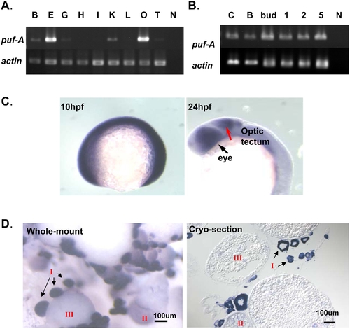
Expression of puf-A in zebrafish using RT-PCR and in situ hybridization. (A) Gene expression in adult tissues of zebrafish was analyzed by RT-PCR and electrophoresis with puf-A primers (upper panel) or actin primers (lower panel, as the internal control). Notation: B, brain; E, eye; G, gill; H, heart; I, intestine; K, head kidney; L, liver; O, ovary; T, testis; N, negative control. (B) The puf-A gene was expressed in various stages of zebrafish embryo. C, cleavage; B, blastula; 1, 1 day post-fertilization (dpf); 2, 2 dpf; 5, 5 dpf; N, negative control. (C) Whole-mount in situ hybridization with puf-A antisense riboprobe on zebrafish embryo. The puf-A expression at 10 h post-fertilization (hpf) (tailbud stage) and 24 hpf (25-somite stage) with lateral overview. The black arrow points to the eye and the red arrow to the optic tectum. (D) Whole-mount and cryo-section in situ hybridization with a puf-A antisense riboprobe in adult ovaries. In adult ovaries, a staging series of oocyte development was characterized by the diameter of various oocytes [9], [10]. Stage I, primary growth follicles (<0.1 mm); stage II, previtellogenic (0.1–0.30 mm); and stage III, vitellogenic (>0.30 mm). |
| Gene: | |
|---|---|
| Fish: | |
| Anatomical Terms: | |
| Stage Range: | 2-cell to Adult |