
Fig. 6
- ID
- ZDB-FIG-090320-69
- Publication
- Holloway et al., 2009 - A novel role for MAPKAPK2 in morphogenesis during zebrafish development
- Other Figures
- All Figure Page
- Back to All Figure Page
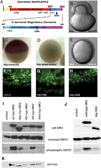
Zebrafish MAPKAPK2 protein schematic, sub-cellular localization, and kinase activity. (A) Top panel: Full length protein of 382 amino acids, with catalytic domain (red) adjacent to the carboxy-terminal regulatory domains (blue, purple). Residues T202, S252, and T315 (*) are phosphorylated by p38 MAPK. K73M is the kinase dead mutation generated. The Q350 residue (black arrowhead) is mutated to a premature stop codon in bbp, thus creating a truncated protein lacking the most carboxy-terminal regulatory domains (purple). Bottom panel: Scale representation of carboxy-terminal regulatory domains, including the auto-inhibitory helix that slightly overlaps an NES, and the NLS that also contains residues required for p38 MAPK docking (p38). (B) bbp embryo injected with kinase-dead full-length myc-tagged MAPKAPK2 (MK2), which does not rescue; shown at 50% epiboly when the constriction initiated. (C,D) Wholemount in situ hybridization of MK2 (bbp) antisense and control sense probes at 50% epiboly in WT embryos. (E) bbp embryo injected with full-length myc-tagged MK2 mRNA at the 1-cell stage is rescued; shown at 75% epiboly. (F–H) Anti-myc antibody staining of WT (bbp/+) embryos expressing WT (F), bbp (G), and a non-phosphorylatable T202A/S252A/T315A triple mutation (H) of myc-tagged MK2 (100 pg mRNA injected). I. Western blot of HeLa cell extracts expressing a vector control (lane 1), full-length myc-tagged MK2 (lane 2), truncated myc-tagged Bbp MK2 (lane 3), full-length kinase dead myc-tagged MK2 (lane 4), T202A/T315A myc-tagged MK2 (lane 5), T202A/S252A/T315A myc-tagged MK2 (lane 6). (J) Western blotting of HeLa cell extracts expressing a vector control (lane 1), full-length untagged MK2 (lane 2), or truncated untagged Bbp MK2 (lane 3). (I, J) HeLa cell extract blots were probed for total MK2 protein (MK2) (top panels), total HSP27 protein (middle panels), and phosphorylated HSP27 protein (bottom panels). (K) Western blot probed with anti-myc of embryo extracts either uninjected (lane 1) or expressing myc-tagged MK2 constructs, as described in I. |