
Fig. 5
- ID
- ZDB-FIG-090220-40
- Publication
- Pei et al., 2009 - Identification of common and unique modifiers of zebrafish midline bifurcation and cyclopia
- Other Figures
- All Figure Page
- Back to All Figure Page
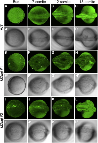
Time-lapse imaging of midline bifurcation formation and recovery. Embryos vitally-stained with fluorescent bodipy ceramide were mounted in soft agarose and incubated at 34 °C except for ∼ 15 minute interruptions at room temperature (∼22 °C) for microphotography, performed every ∼2 h. (A–D) Midline formation in heated WT embryos. Heated WT embryos developed well-defined dorsal midlines during the recording. (E–L) Midline formation in two heated MZsqt embryos. (E–H) MZsqt #1 embryos formed a large bifurcation, first visible at the 7-somite stage. (I–L) MZsqt #2 embryos displayed a small bifurcation, first visible at the bud stage (I), which recovered by the 12-somite stage (K). Because embryos were incubated and photographed at temperatures (34 °C and 22 °C) causing distinct developmental rates, the indicated stages are based on morphology, rather than time. Apostrophes (') indicate bright field images of the same specimen. Dorsal views are given throughout, with the anterior to the right. |
| Fish: | |
|---|---|
| Condition: | |
| Observed In: | |
| Stage Range: | 5-9 somites to 14-19 somites |
Reprinted from Developmental Biology, 326(1), Pei, W., and Feldman, B., Identification of common and unique modifiers of zebrafish midline bifurcation and cyclopia, 201-211, Copyright (2009) with permission from Elsevier. Full text @ Dev. Biol.