
Fig. 5
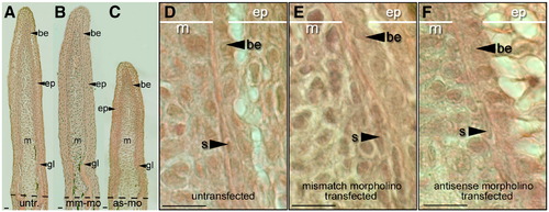
Tissues of the early regenerating fin are still present in morpholino-transfected regenerating fins. (A) Coronal sections through a regenerating caudal fin from a wild-type zebrafish 3 dpa. The regenerative outgrowth of the fin stained with Hematoxylin and Eosin: the epidermis (ep), basal epithelium (be), mesenchyme (m) and growing lepidotrichia (gl). (B) Section through fin lobes transfected with mismatch control morpholinos. (C) Fins transfected with antisense morpholinos. (Dashed lines show amputation plane.) (D) High magnification of coronal section through untransfected regenerating fins show all cell types (s: scleroblasts). (E) High magnification of regenerate transfected with mismatch control morpholino. (F) High magnification of a regenerate transfected with antisense morpholino. Scale bars equal 100 μm (A–C) and 25 μm (D–F). |
| Fish: | |
|---|---|
| Condition: | |
| Knockdown Reagent: | |
| Observed In: | |
| Stage: | Adult |
Reprinted from Developmental Biology, 325(2), Kizil, C., Otto, G.W., Geisler, R., Nüsslein-Volhard, C., and Antos, C.L., Simplet controls cell proliferation and gene transcription during zebrafish caudal fin regeneration, 329-340, Copyright (2009) with permission from Elsevier. Full text @ Dev. Biol.