FIGURE

Fig. 6

ID
ZDB-FIG-090127-25
Publication
Targoff et al., 2008 - Nkx genes regulate heart tube extension and exert differential effects on ventricular and atrial cell number
Other Figures
All Figure Page
Back to All Figure Page
Fig. 6

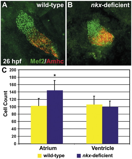
nkx genes limit atrial cell number during heart tube assembly. (A,B) Immunofluorescence indicates nuclear localization of Mef2 (green) throughout the heart tube, facilitating counting of differentiated cardiomyocytes at 26 hpf. Atrial cells are indicated by the anti-Amhc antibody, S46 (red). Hearts are flattened with a cover slip to improve visualization of cardiomyocyte nuclei. (C) Quantification of cardiomyocyte nuclei in wild-type (n = 25) and nkx-deficient (n = 11) embryos reveals a statistically significant increase in atrial cell number in nkx-deficient embryos (p < 0.001, Student's t-test) and no significant difference in ventricular cell number. Bar graph indicates mean and standard error of each data set; asterisk indicates statistically significant difference from wild-type.

Expression Data
Antibodies:
Fish:
Knockdown Reagents:
Anatomical Term:
Stage: Prim-5

Expression Detail
Antibody Labeling
Phenotype Data
Fish:
Knockdown Reagents:
Observed In:
Stage: Prim-5

Phenotype Detail
Acknowledgments
This image is the copyrighted work of the attributed author or publisher, and ZFIN has permission only to display this image to its users. Additional permissions should be obtained from the applicable author or publisher of the image.

Reprinted from Developmental Biology, 322(2), Targoff, K.L., Schell, T., and Yelon, D., Nkx genes regulate heart tube extension and exert differential effects on ventricular and atrial cell number, 314-321, Copyright (2008) with permission from Elsevier. Full text @ Dev. Biol.