|
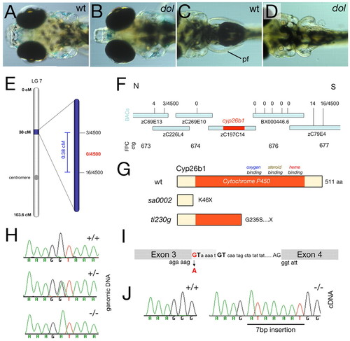
dolphin corresponds to cyp26b1. (A-D) Live zebrafish larvae at 120 hpf. Dorsal views of head (A,B) and on pectoral fins (pf; C,D). wt, wild type; dol, dolphin allele ti230g. (E)The dol-bearing interval on chromosome 7. (F) Interval-spanning BAC contig, with indicated recombinations in 4500 meioses and showing the location of cyp26b1 gene. (G) Schematic of wild-type and mutant zebrafish Cyp26b1 proteins. (H) Sequencing profiles of cyp26b1 exon 3-intron junction from genomic DNA of wild type (+/+), heterozygous (+/-) and homozygous (-/-) ti230g mutants. (I) Schematic of exon 3-intron junction of cyp26b1. The mutated G in the splice-donor site of the ti230g allele is in red, the internal GT used in the mutant is in bold, and the 7 bp insert in mutant cDNA is in light gray. (J) Sequencing profiles of cyp26b1 exon 3-exon 4 junction from cDNA of wild type and ti230g mutants. The inserted sequence is underlined.
|